Pectinate Muscles And Trabeculae Carneae . The walls of the ventricle are lined with trabeculae carneae, ridges of cardiac muscle covered by endocardium (figure \(\pageindex{11}\)) that. There are two papillary muscles present which attach. Pectinate muscles differ to the muscular ridges seen in the walls of the ventricles, termed trabeculae carneae. The walls of the ventricle are lined with trabeculae carneae, ridges of cardiac muscle covered by endocardium (figure \(\pageindex{11}\)) that increase the surface area of the. The left atrial appendage is much smaller than that of the right atrium and is not marked by any specific crista internally or sulcus externally. The walls of the inflow portion of the left ventricle are lined by trabeculae carneae, as described with the right ventricle.
from www.numerade.com
Pectinate muscles differ to the muscular ridges seen in the walls of the ventricles, termed trabeculae carneae. The walls of the inflow portion of the left ventricle are lined by trabeculae carneae, as described with the right ventricle. The left atrial appendage is much smaller than that of the right atrium and is not marked by any specific crista internally or sulcus externally. The walls of the ventricle are lined with trabeculae carneae, ridges of cardiac muscle covered by endocardium (figure \(\pageindex{11}\)) that. The walls of the ventricle are lined with trabeculae carneae, ridges of cardiac muscle covered by endocardium (figure \(\pageindex{11}\)) that increase the surface area of the. There are two papillary muscles present which attach.
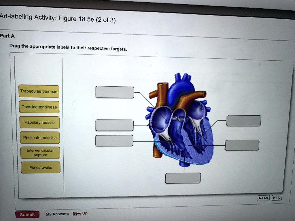
SOLVED Artlabeling ActivityFigure 18.5e (2 of 3 Part A Drag the
Pectinate Muscles And Trabeculae Carneae The walls of the ventricle are lined with trabeculae carneae, ridges of cardiac muscle covered by endocardium (figure \(\pageindex{11}\)) that. The walls of the inflow portion of the left ventricle are lined by trabeculae carneae, as described with the right ventricle. The walls of the ventricle are lined with trabeculae carneae, ridges of cardiac muscle covered by endocardium (figure \(\pageindex{11}\)) that increase the surface area of the. The left atrial appendage is much smaller than that of the right atrium and is not marked by any specific crista internally or sulcus externally. Pectinate muscles differ to the muscular ridges seen in the walls of the ventricles, termed trabeculae carneae. The walls of the ventricle are lined with trabeculae carneae, ridges of cardiac muscle covered by endocardium (figure \(\pageindex{11}\)) that. There are two papillary muscles present which attach.
From www.numerade.com
Complete the labeling activity. Drag the appropriate labels to their Pectinate Muscles And Trabeculae Carneae The walls of the inflow portion of the left ventricle are lined by trabeculae carneae, as described with the right ventricle. The left atrial appendage is much smaller than that of the right atrium and is not marked by any specific crista internally or sulcus externally. There are two papillary muscles present which attach. The walls of the ventricle are. Pectinate Muscles And Trabeculae Carneae.
From www.slideserve.com
PPT HEART PowerPoint Presentation, free download ID463693 Pectinate Muscles And Trabeculae Carneae Pectinate muscles differ to the muscular ridges seen in the walls of the ventricles, termed trabeculae carneae. The left atrial appendage is much smaller than that of the right atrium and is not marked by any specific crista internally or sulcus externally. The walls of the ventricle are lined with trabeculae carneae, ridges of cardiac muscle covered by endocardium (figure. Pectinate Muscles And Trabeculae Carneae.
From www.slideserve.com
PPT Tissues of the Heart PowerPoint Presentation, free download ID Pectinate Muscles And Trabeculae Carneae There are two papillary muscles present which attach. The walls of the inflow portion of the left ventricle are lined by trabeculae carneae, as described with the right ventricle. The walls of the ventricle are lined with trabeculae carneae, ridges of cardiac muscle covered by endocardium (figure \(\pageindex{11}\)) that. The walls of the ventricle are lined with trabeculae carneae, ridges. Pectinate Muscles And Trabeculae Carneae.
From www.chegg.com
Solved Which structure is highlighted? 1 O Multiple Choice Pectinate Muscles And Trabeculae Carneae Pectinate muscles differ to the muscular ridges seen in the walls of the ventricles, termed trabeculae carneae. There are two papillary muscles present which attach. The walls of the ventricle are lined with trabeculae carneae, ridges of cardiac muscle covered by endocardium (figure \(\pageindex{11}\)) that increase the surface area of the. The walls of the ventricle are lined with trabeculae. Pectinate Muscles And Trabeculae Carneae.
From teachmeanatomy.info
Chambers of the Heart Atria Ventricles TeachMeAnatomy Pectinate Muscles And Trabeculae Carneae The walls of the inflow portion of the left ventricle are lined by trabeculae carneae, as described with the right ventricle. There are two papillary muscles present which attach. Pectinate muscles differ to the muscular ridges seen in the walls of the ventricles, termed trabeculae carneae. The walls of the ventricle are lined with trabeculae carneae, ridges of cardiac muscle. Pectinate Muscles And Trabeculae Carneae.
From www.slideserve.com
PPT Ex. 41 Structure of the Heart PowerPoint Presentation, free Pectinate Muscles And Trabeculae Carneae The walls of the ventricle are lined with trabeculae carneae, ridges of cardiac muscle covered by endocardium (figure \(\pageindex{11}\)) that increase the surface area of the. The left atrial appendage is much smaller than that of the right atrium and is not marked by any specific crista internally or sulcus externally. The walls of the inflow portion of the left. Pectinate Muscles And Trabeculae Carneae.
From www.flickr.com
Trabeculae carneae The Anatomy of the Heart Visual Atlas… Flickr Pectinate Muscles And Trabeculae Carneae The walls of the ventricle are lined with trabeculae carneae, ridges of cardiac muscle covered by endocardium (figure \(\pageindex{11}\)) that. The walls of the inflow portion of the left ventricle are lined by trabeculae carneae, as described with the right ventricle. There are two papillary muscles present which attach. Pectinate muscles differ to the muscular ridges seen in the walls. Pectinate Muscles And Trabeculae Carneae.
From www.numerade.com
SOLVED Which structure is highlighted? Multiple Choice A. trabeculae Pectinate Muscles And Trabeculae Carneae Pectinate muscles differ to the muscular ridges seen in the walls of the ventricles, termed trabeculae carneae. The walls of the ventricle are lined with trabeculae carneae, ridges of cardiac muscle covered by endocardium (figure \(\pageindex{11}\)) that. The left atrial appendage is much smaller than that of the right atrium and is not marked by any specific crista internally or. Pectinate Muscles And Trabeculae Carneae.
From www.chegg.com
Solved Which structure is highlighted? trabeculae carne Pectinate Muscles And Trabeculae Carneae The walls of the ventricle are lined with trabeculae carneae, ridges of cardiac muscle covered by endocardium (figure \(\pageindex{11}\)) that increase the surface area of the. There are two papillary muscles present which attach. Pectinate muscles differ to the muscular ridges seen in the walls of the ventricles, termed trabeculae carneae. The left atrial appendage is much smaller than that. Pectinate Muscles And Trabeculae Carneae.
From www.chegg.com
Solved Apex of the heart Cusp of tricuspid valve Right Pectinate Muscles And Trabeculae Carneae The walls of the ventricle are lined with trabeculae carneae, ridges of cardiac muscle covered by endocardium (figure \(\pageindex{11}\)) that increase the surface area of the. The left atrial appendage is much smaller than that of the right atrium and is not marked by any specific crista internally or sulcus externally. There are two papillary muscles present which attach. Pectinate. Pectinate Muscles And Trabeculae Carneae.
From www.chegg.com
Solved Question 24 options Pectinate Muscle Chordae Pectinate Muscles And Trabeculae Carneae The walls of the ventricle are lined with trabeculae carneae, ridges of cardiac muscle covered by endocardium (figure \(\pageindex{11}\)) that increase the surface area of the. There are two papillary muscles present which attach. The walls of the inflow portion of the left ventricle are lined by trabeculae carneae, as described with the right ventricle. The walls of the ventricle. Pectinate Muscles And Trabeculae Carneae.
From www.numerade.com
SOLVED 6. The muscles that attach to tendinous cords (chordae Pectinate Muscles And Trabeculae Carneae There are two papillary muscles present which attach. The walls of the ventricle are lined with trabeculae carneae, ridges of cardiac muscle covered by endocardium (figure \(\pageindex{11}\)) that. Pectinate muscles differ to the muscular ridges seen in the walls of the ventricles, termed trabeculae carneae. The left atrial appendage is much smaller than that of the right atrium and is. Pectinate Muscles And Trabeculae Carneae.
From www.chegg.com
Solved Chordae tendineae Trabeculae carneae Pectinate Pectinate Muscles And Trabeculae Carneae The walls of the inflow portion of the left ventricle are lined by trabeculae carneae, as described with the right ventricle. There are two papillary muscles present which attach. The walls of the ventricle are lined with trabeculae carneae, ridges of cardiac muscle covered by endocardium (figure \(\pageindex{11}\)) that. The walls of the ventricle are lined with trabeculae carneae, ridges. Pectinate Muscles And Trabeculae Carneae.
From www.chegg.com
Solved Drag the structure names to the appropriate locations Pectinate Muscles And Trabeculae Carneae There are two papillary muscles present which attach. The walls of the ventricle are lined with trabeculae carneae, ridges of cardiac muscle covered by endocardium (figure \(\pageindex{11}\)) that increase the surface area of the. The left atrial appendage is much smaller than that of the right atrium and is not marked by any specific crista internally or sulcus externally. Pectinate. Pectinate Muscles And Trabeculae Carneae.
From www.slideserve.com
PPT Ex. 41 Structure of the Heart PowerPoint Presentation, free Pectinate Muscles And Trabeculae Carneae The walls of the ventricle are lined with trabeculae carneae, ridges of cardiac muscle covered by endocardium (figure \(\pageindex{11}\)) that. The walls of the inflow portion of the left ventricle are lined by trabeculae carneae, as described with the right ventricle. There are two papillary muscles present which attach. The walls of the ventricle are lined with trabeculae carneae, ridges. Pectinate Muscles And Trabeculae Carneae.
From www.animalia-life.club
Trabeculae Carneae Vs Papillary Muscle Pectinate Muscles And Trabeculae Carneae Pectinate muscles differ to the muscular ridges seen in the walls of the ventricles, termed trabeculae carneae. The walls of the ventricle are lined with trabeculae carneae, ridges of cardiac muscle covered by endocardium (figure \(\pageindex{11}\)) that increase the surface area of the. The left atrial appendage is much smaller than that of the right atrium and is not marked. Pectinate Muscles And Trabeculae Carneae.
From www.numerade.com
SOLVED Artlabeling ActivityFigure 18.5e (2 of 3 Part A Drag the Pectinate Muscles And Trabeculae Carneae The left atrial appendage is much smaller than that of the right atrium and is not marked by any specific crista internally or sulcus externally. The walls of the inflow portion of the left ventricle are lined by trabeculae carneae, as described with the right ventricle. Pectinate muscles differ to the muscular ridges seen in the walls of the ventricles,. Pectinate Muscles And Trabeculae Carneae.
From quizlet.com
Models 10 Diagram Quizlet Pectinate Muscles And Trabeculae Carneae The walls of the ventricle are lined with trabeculae carneae, ridges of cardiac muscle covered by endocardium (figure \(\pageindex{11}\)) that. The walls of the ventricle are lined with trabeculae carneae, ridges of cardiac muscle covered by endocardium (figure \(\pageindex{11}\)) that increase the surface area of the. The left atrial appendage is much smaller than that of the right atrium and. Pectinate Muscles And Trabeculae Carneae.